on July 17, 2007
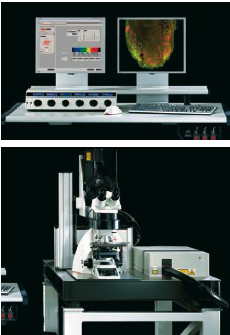
The Swiss National Fund agreed recently to fund half of the cost of the purchase of a highly sophisticated microscope.The generosity of a local foundation has agreed to cover the other half of the 850’000.- cost of the instrument.This latest step in improving the infrastructure of the IRB underlines the strategic importance of improving the instruments available to IRB scientists enabling their ambitious programs. Under its R’EQUIP Programme (Research Equipment), the Swiss National Science Foundation (SNSF) supports the purchase, development and moderniza-tion (upgrading) of research equipment, which is essential for the launch of new research facilities.
In many research areas success is dependent on the availability of top quality equipment. In recent years, investment in such research equipment at Swiss higher education institutions has declined to a relatively low level.The SNSF has set itself the objective with its R’EQUIP Programme of forestalling this trend before any deficits become evident in the national research infrastructure.
Advanced imaging technology has become essential for timely, competitive biomedical research and the lack of such equipment in Ticino has blocked development of numerous projects.The successful application to the SNF for funding demonstrates how 6 independent research projects of relevance to Immunology, Hematology,AIDS, and Alzheimer’s disease, each from a different SNF-funded Groups,will be advanced by access to the new microscope equipment.These projects are but the “tip of the iceberg”, when one considers the enormous potential utilization of state-of-the-art microscopy by investigators from IRB and collaborating institutions. Basic concept: the principle of confocal imaging was patented by Marvin Minsky in 1957. In a conventional (i.e., wide-field) fluorescence microscope, the entire specimen is flooded with light from a light source. In contrast, a confocal microscope uses point illumination and a pinhole in an optically conjugate plane in front of the detector to eliminate out-of-focus information. Only the light within the focal plane can be detected, so the image quality is much better than that of wide-field images. As only one point is illuminated at a time in confocal microscopy, 2D or 3D imaging requires scanning over a regular raster (i.e. a rectangular pattern of parallel scanning lines) in the specimen.
The thickness of the focal plane is defined mostly by the square of the numerical aperture of the objective lens, and also by the optical properties of the specimen and the ambient index of refraction.